Human Bone Gallery - Deficiency diseases

Rickets of the legs

Rickets

Site: Jarrow monastic site, Tyne and Wear.
Period: Late Saxon to post-medieval.
Excavator: Prof. R.J. Cramp, Durham University.
Published: forthcoming.
Skeleton: child, 4 years.

Catalogue entry: The left femur has extensive lateral bowing in its distal two-thirds with wide splaying of its distal end. The right femur is deformed in the same way, but less so. The left tibia and fibula are also much deformed, being bowed laterally from just below the head. The right tibia and fibula are somewhat less affected although also deformed. It is likely that this child was an early sufferer of rickets.

Rickets is the result of vitamin D deficiency and is more common in post-medieval and early modern urban industrial populations. The avitaminosis may be attributable to malnutrition, but is also linked with lack of exposure to sunlight. Disease may be a contributory factor, and in this case there were other pathological changes which may indicate an infection or inflammation of the bone.

Cribra orbitalia

Cribra orbitalia

Site: Jarrow monastic site, Tyne and Wear.
Period: Late Saxon to post-medieval.
Excavator: Prof. R.J. Cramp, Durham University.
Published: forthcoming.
Skeleton: child, 2-3 years.

Catalogue entry: Both orbits are affected with cribra orbitalia. The right side is cribriotic (2nd degree) and the left trabecular (3rd degree).

Cribra orbitalia is a pathological lesion of the roof of the eye socket (orbit) which has been associated with iron deficiency anaemia. It causes pitting, porosity of the smooth bone in the orbit, and sometimes new thickened bone growth. It is particularly prevalent in children, and as with this child, the degree of the condition is often greater in the very young.

Enamel hypoplasia

Enamel hypoplasia

Site: The Hirsel churchyard, Coldstream, Scotland.
Period: Medieval.
Excavator: Prof. R.J. Cramp, Durham University.
Published: forthcoming (Historic Scotland?).
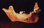
Skeleton: child, c.7 years.

Catalogue entry: A disarticulated mandible of a child aged c.7 years, found with an older child (c.12 years). The first adult molars are fully erupted, but the seconds are unerupted. The adult incisors are almost fully erupted. Both juvenile canines have been lost post-mortem, and only one of the deciduous molars, the left second, is still in situ.

The teeth have gross hypoplastic lesions; the lower mesial incisors and the lower first molars each have three large ridges at the cementum-enamel junction.