Introduction to Human Bones
Why excavate human bones?
Archaeologists often have to excavate cemeteries and individual burials to 'clear' a site for development. If archaeologists didn't do this work, other contractors would remove the burials.
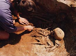
Bones provide a huge amount of information about people in the past. Medieval and earlier burials are usually anonymous, but they provide information on ordinary people which is rarely found in historical studies.
Burials are a good source of information about beliefs, ritual behaviour and treatment of the dead.
What can we learn from studying them?
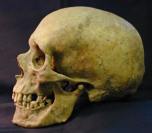
Archaeologists study people in the past, so study of the physical remains of actual people is fundamental.
The study of human remains adds another dimension to the past which study of the material culture cannot usually provide, namely the health and physique of a population.
The basic requirements of the archaeologist dictate the major aspects of work carried out by the archaeological human bone specialist: age and sex structure of a population; physical size and appearance; general stresses and strains of daily life; and diseases in the past.
The techniques of analysing bones
Introduction
The following is an introduction to the work of the archaeological human bone specialist, osteoarchaeologist, or physical/biological anthropologist. The general techniques are used widely by both archaeological and forensic specialists in human remains. Further and more detailed information is available in the books referenced at the end of this page.
Age
The most basic distinction is between child and adult. Although clearly children are generally smaller than adults, this is not a reliable method of distinction beyond the age of c.12 years.
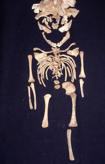
The ends of the long bones and parts of some other bones are detached in children's skeletons. This allows the bones to grow. These detached parts are known as epiphyses, and they all fuse at different ages, from c.15 years onwards. The stage of fusion of the bones can therefore be used to age an adolescent. Before this, the most reliable method is to study the developmental stage of the teeth. As a rough guide, the first permanent molar erupts at the age of six years, the second at twelve years, and the third at c.18-21 years - you need to know the difference between milk molars and permanent molars first though! If the teeth are not present then the long bone lengths are also a good indicator of age in younger children.
Ageing skeletons becomes more difficult as the individual gets older. Once all the bones have fused there is really no reliable simple method to use. Tooth wear gives some indication, but only if members of the group being studied were eating a fairly coarse diet. Other methods of ageing, such as changes to the face of the pubic symphysis (the joint at the front of the pelvis) or cranial suture closure, have largely been discredited in recent years. The best we can do is to place skeletons into simple age categories - young, middle-aged or old.
Sex
It is not generally possible to sex the skeleton of a child, unless it has passed puberty and is starting to show some of the characteristics appropriate to its sex. Adults are more easy to deal with, as long as you remember that humans are continuously variable and there is not always an easy distinction to be made between a small man and a large woman.
The best indicators of sex in the skeleton are to be found in the pelvis. This is because one of the major biological differences between men and women, that of having babies, largely determines the shape of that part of the body. A woman's pelvis is wide and bowl-shaped whilst a man's is tall and narrow. As the pelvis is made up of three bones (two innominates and the sacrum), this can be seen in the width of the sacrum, the sciatic notch and the sub-pubic angle, and sex can usually be determined even if part of the pelvis is destroyed.
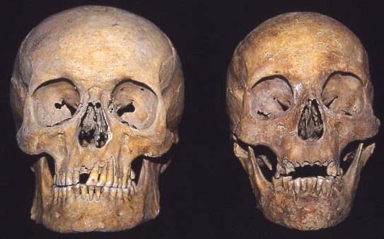
The next best indicator of sex is the skull. The face shows subtle differences in the shape of the eye sockets (orbits), the angle of the forehead is more vertical in women, the brow ridges and mastoid processes are smaller, and the muscle markings are less robust. In fact this is the main criterion for determining sex if only the long bones and a few fragments of skull remain - as a general rule men tend to be more physically robust than women.
Metric analysis
Metric analysis involves using standard measurements defined in the 19th and 20th centuries for recording the size of various attributes of the human skeleton. For general purposes, these consist of the length, breadth and height of the skull and facial bones, and the lengths of long bones. Some measurements are used to calculate indices which show the relationship between different dimensions. For example, the cranial index is calculated to show the relationship between length and breadth of the cranial vault. It can then be divided into groups: narrow-, medium- and broad-headed (dolicho-, meso- and brachycranial respectively).
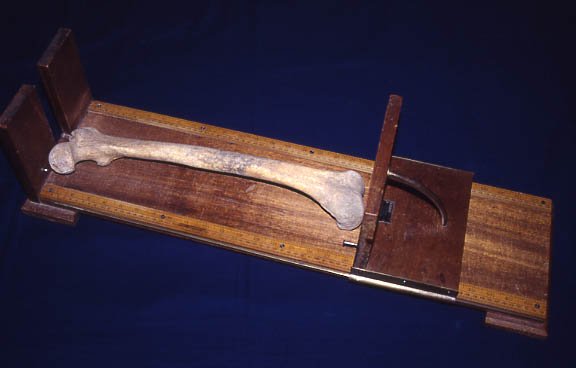
Tools used are sliding and spreading callipers, and an osteometric board to measure long bone lengths.
Measurement forms are used by many specialists to ensure standardisation of the list used for each skeleton. A typical form may have 27 cranial measurements (although there are many more — up to 500!) and 8 indices, 10 mandibular measurements and 23 post-cranial measurements. Most of these are defined in the standard British text, Brothwell's Digging Up Bones.
Stature equations were calculated from skeletons of recent Americans whose living stature was known. Whilst they may slightly overestimate for British and Anglo-Saxon groups, proportions of the limbs in these groups suggest that they are similar enough for general use.
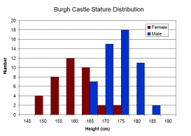
The main purpose of measurement is comparison. Stature may be compared between groups or sexes. Usually this shows some overlap between males and females, as would be expected. The normal average in most Roman to medieval groups was around 5' 7" for males and 5' 3" for females. Changes in the cranial index occur through time; there is a general increase from the Saxon to the Medieval period, with rounder heads in the latter. A similar change is well-documented from the Neolithic to the Bronze Age. In the past, this has been seen as evidence for a change in population, perhaps due to invasion, but other interpretations are possible. For example, climate may have an effect on head shape.
Statistical tests can be used for comparison of measurements between sites.
Non-metric traits
Non-metric traits, also known as discontinuous or genetic traits, are anomalies in the skeleton which are recorded on a present or absent basis. Usually they are too small to measure. Their presence or absence can tell us about family relationships within a group, and they can also be used for comparisons between groups.
Unfortunately, we do not know the genetics of all the traits which we record, but a number are certainly inherited. A few may be more related to physical robustness and have shown a marked sex difference in some groups.
Examples of non-metric traits include the following:
Metopism — the frontal bone of the skull is divided into two halves in the newborn infant, and eventually fuses together between the ages of 2 and 6 years. Sometimes fusion does not take place, and a suture, the metopic suture, remains in place of the original division. This trait occurs at a rate of 5-10% in most archaeological populations and has proved very useful for identifying family groups.
Wormian bones — these are extra bones which occur between the plates of the skull vault. They are particularly common in the lambdoid suture, but also occur in the sagittal and coronal sutures, and to a lesser extent in other cranial vault sutures. When they occur at joints of the sutures they have special names - the inca bone occurs at the junction of the lambdoid and sagittal sutures, whilst the bregmatic bone occurs at the junction of the sagittal and coronal sutures.
Tori — a torus may occur in the centre line of the palate, on either side of the maxilla, on either side of the mandible, or in the ears. These anomalies are bony protuberances which appear to be genetic in origin. Mandibular tori are known to be particularly common in some groups of people, particularly the Inuit. Auditory tori have recently been associated with swimming.
Other cranial traits include the presence or absence of certain holes (foramina), particularly those on the parietal, and around the eye sockets. Some of these traits are so common that it is more unusual not to find them.
A few traits are recorded in the rest of the skeleton, although there is less agreement about which of these have a congenital element. The following are some of the most common traits:
Septal aperture — a small hole sometimes forms at the elbow joint in the distal end of the humerus. This tends to occur more often in female skeletons and may be related to robusticity. However, it has a widely varying prevalence and it is useful for comparisons between groups.
Femoral third trochanter — 'trochanter' is the anatomical term for one of three processes which form on the femur. The greater and lesser trochanters are present in normal anatomy, but the third trochanter is unusual and may be genetically determined. It forms at the back of the bone just below the hip joint, near the attachment for the gluteal muscle. In some groups it is more common in men, and may be related to robusticity. It should not be confused with the gluteal tuberosity, a roughened area of bone which is a trait of robusticity.
Vertebral traits — there are many possibilities for variety within the human spine. Sometimes there are more or less vertebrae than normal, for example 13 thoracic vertebrae instead of the usual twelve. Sometimes ribs may form on the last cervical or first lumbar vertebrae, or the vertebral arch may not fuse to the body.
Dental analysis
Dental analysis involves recording the state of eruption, tooth loss (before or after death) and dental pathology for each tooth in the mouth. This data can then be used to calculate the prevalence of dental disease within a group of skeletons.
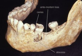
The most common findings include dental caries, abscesses and ante-mortem tooth loss (teeth lost in life). Unerupted or impacted third molars (wisdom teeth) are also common. Many individuals were affected with dental calculus (tartar), and some have a condition known as enamel hypoplasia (lines or pits in the enamel related to malnutrition or illness as a child). Sometimes there may be retention of a milk tooth amongst otherwise adult dentition, overcrowding, abnormally small teeth, or other unusual anomalies.
Study of the teeth is useful in showing changes in dental health, particularly increasing rates of caries and abscesses from the Saxon to the Medieval period. Different rates of dental disease can be seen in different age groups, with the oldest members of a population generally being most affected by dental disease and tooth loss. Dietary information may be obtained from the teeth — e.g. wear suggests heavily gritted food. Finally, interpretations regarding dental health care can be made, e.g. in earlier groups the presence of calculus suggests poor oral hygiene, whilst later groups may have false teeth and fillings.
Palaeopathology
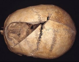
Palaeopathology is the study of ancient diseases. A number of sources can be used for this including documents, art, and medical instruments. Here, we are concerned with the evidence found in the skeleton, although some palaeopathologists are able to use soft tissue evidence from mummies and bog bodies. Unfortunately for bone specialists, most diseases do not reach the bone, so we have no evidence for illnesses such as measles, smallpox, plague, influenza, and many other diseases which we assume to have been present in the past. Descriptions of illnesses in Roman and later documents can be used as a guide, but different names were used and the symptoms described are not always easy to interpret.
Palaeopathologists tend to categorise diseases by probable cause, although diagnosis is often difficult because many pathological changes in bone have a similar appearance. I use the following categories when writing a report:
- Congenital diseases: These include mild conditions such as spina bifida occulta of the sacrum, cervical ribs, extra vertebrae, etc.
- Arthropathies and degenerative disease: These are the most common diseases to be found in skeletal material, and include osteoarthritis, osteoporosis, and general changes associated with the ageing process.
- General spinal pathology: The spine is a common area for stress injuries and pain, mainly due to our bipedalism. A number of common pathological changes are found, such as Schmorl's nodes, and anterior epiphyseal dysplasia.
- Metabolic and nutritional disorders: Bone is easily affected by poor nutrition, and produces changes related to iron deficiency anaemia (cribra orbitalia) and Vitamin D deficiency (rickets, osteomalacia). Other changes which may be related to malnutriton or illness can be seen in the teeth (enamel hypoplasia) and the long bones (Harris lines).
- Circulatory disturbances: If the blood supply to an area of bone is cut off, that area of the skeleton will die and may be resorbed. Osteochondritis dissecans is a relatively common disease which falls into this category and which tends to be associated with physical trauma.
- Infectious diseases: Not many infections reach the bone. Acute diseases such as the plague and smallpox tend to kill or cure very quickly and do not have time to affect the skeleton. Chronic illnesses such as tuberculosis, leprosy and syphilis can be recognised in human remains. Inflammatory changes such as periostitis (affecting the outer layer of a bone) and sinusitis (affecting the sinuses) may also be caused by infections, although other causes are also possible.
- Trauma: This category can include anything from a simple sprain or bruise to the most spectacular of head wounds. Sometimes the injuries may be so bad that the individual died from them, in which case they may not be easily recognisable, but often fractures, cuts and surgical interventions have healed and can be identified.
- Neoplasms: This category covers diseases associated with the formation of tumours, although generally those seen in the bone are benign 'warty' lesions. Malignant tumours are rare in archaeological bones.
- Miscellaneous lesions: This category covers any pathologies which do not fit easily into the categories above, or which cannot be identified.
Further reading
Brothwell, D. 1981 Digging Up Bones. London: BM(NH)/OUP.
Chamberlain, A. 1994 Human Remains. London: British Museum Press.
Mays, S. 1998 The Archaeology of Human Bones. London: Routledge.
Stirland, A. 1999 Human Bones in Archaeology. 2nd ed. Aylesbury: Shire Publications.
Roberts, C. 2009 Human Remains in Archaeology: a Handbook. York: Council for British Archaeology.
Waldron, T. 2001 Shadows in the Soil. Human bones and archaeology. Stroud: Tempus.