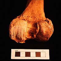
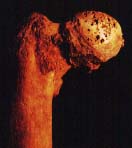
Osteoarthritis: knee
Site: Burgh Castle, Norfolk.
Period: Middle Saxon cemetery in a Roman fort.
Excavator: Charles Green.
Published: East Anglian Archaeology (bone report in Caister volume).
Skeleton: ?male in middle or old age.
Catalogue entry: A disarticulated right femur showing gross osteoarthritic changes to the distal end. The medial condyle is flattened and striated, with thickening and eburnation of the joint surface. Both condyles are edged with large osteophytes, and there is pitting on the surface of the lateral condyle.
Changes of this kind can only occur if the surfaces of the bone joints are in contact. At this stage of the disease, the cartilaginous pad is destroyed and pain is severe. The state of the knee joint in this case indicates that the individual continued to walk on it and he must have suffered agony when moving. |