Human Bone Gallery - Developmental/congenital conditions
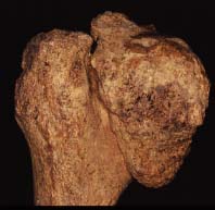 |
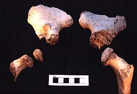
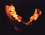
Slipped femoral capital epiphysisSite: St. Saviour's Hospital, Bury St. Edmunds. Catalogue entry: The left acetabulum of this individual is very wide (57mm in diameter, compared with 48mm on the right side) with a sclerotic floor, eburnation on the superior surface, and very large osteophytes around the rim. The head of the left femur is surrounded by large bony outgrowths. The proximal epiphysis has slipped and is positioned, and fused, on the medio-dorsal surface of the neck, which shows no sign of fracture (although it is partly obscured by new bone growth over the anterior surface). The end of the metaphysis does not appear to have been remodelled, and the lumpy appearance normally associated with the unfused epiphysis is still present in part (although there is some post-mortem erosion). The shape of the head is deformed and curves down to a point at the inferior side. Marked eburnation has occurred on the superior surface, corresponding to that seen in the acetabulum. The proximal half of the shaft of the bone appears slightly remodelled to compensate for the obvious difficulty in walking imposed by this condition, and the bone is slightly narrower medio-laterally than antero-posteriorly. There is slight lipping of the distal condyles and linea aspera, but this is not as marked as that seen on the right femur. This has lipping of the borders of the condyles and a proliferation of bone along the linea aspera and greater and lesser trochanters (which may be related to DISH seen elsewhere in the skeleton). There is eburnation of the lateral condyle with corresponding lesions on the right patella, which is also lipped around the borders. The right femur appears slightly twisted, the linea aspera in the proximal half running almost along the medio-lateral border. The proximal halves of both tibiae are slightly curved towards the lateral side. The heads of all proximal phalanges of the right foot are slightly deformed, and both ends of the second middle phalanx are deformed with osteophyte formation. There is slight eburnation and pitting on the superior lateral surface of the left first metatarsal head. There was evidence of ischial bursitis of the left ischial tuberosity (the right was unaffected), with new bone growth over the surface giving a craggy appearance. The facet for the radius on the right scaphoid has two small patches of eburnation (there is no corresponding lesion on the radius), and there is eburnation on both surfaces of the joint between the scaphoid and the trapezoid. No other bones of the right hand or wrist were affected. For interpretation of these pathological conditions, see the report. |